Back Of Skull Anatomy - Neck Muscles And Other Soft Tissues : Skull reshaping is done on any of the structures that lie above the face.
byAdmin-
0
Back Of Skull Anatomy - Neck Muscles And Other Soft Tissues : Skull reshaping is done on any of the structures that lie above the face.. The occipital bone is located on the back of the cranium and includes. Skull bones aren't fused together at birth. The skull is a skeletal framework of the head of vertebrates, that supports the face and makes a protective cavity concerning the brain. The cranium and the mandible. The neurocranium consists of the frontal, the ethmoid, the sphenoid, the occipital, and the paired temporal and parietal bones.
Looking at it from the inside it can be subdivided into. We monitor our sites and will resolve this issue as soon as possible. Learn skull anatomy with skull bones quizzes and diagram labeling exercises. The cranium (skull) is the skeletal structure of the head that supports the face and protects the brain. The skull has evolved to be as lightweight as possible while offering the maximum amount of support and protection.
The Skull Anatomy And Physiology from philschatz.com The cranium (skull) is the skeletal structure of the head that supports the face and protects the brain. The bbc is not responsible for the content of external websites. It offers protection to the brain, eye balls, inner ears, and nasal passages. Skull, skeletal framework of the head of vertebrates, composed of bones or cartilage, which form a unit that protects the brain and some sense organs. A thorough description is beyond the. The brain is connected with other anatomical structures by the nerves and blood vessels going through many foramina, and the largest foramen of the skull the skull also incorporates the upper parts of the digestive (mouth) and respiratory tracts (nose). The frontal, parietal, temporal and occipital bones are joined at the cranial sutures. Inside the skull, it forms the anterior cranial fossa, which contains the frontal lobes of the cerebrum.
Inside the skull, it forms the anterior cranial fossa, which contains the frontal lobes of the cerebrum.
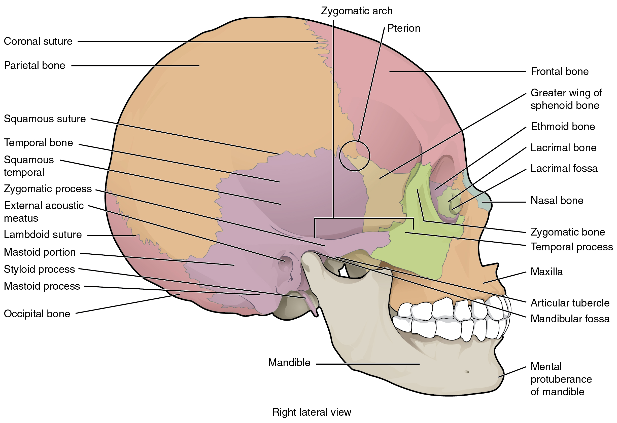
Frontal bone supraorbital rim temporal bone nasal bone zygoma maxilla inferior concha nasal spine mandible glabella greater wing of sphenoid lesser wing of sphenoid optic canal middle concha infraorbital foramen styloid process nasal septum mental foramen. Skull reshaping is done on any of the structures that lie above the face. The skull includes the upper jaw and the cranium. The skull has a single occipital condyle.7 the skull consists of five major bones: They don't move and united into a single unit. Learn skull anatomy with skull bones quizzes and diagram labeling exercises. • it has the supraorbital foramen, where the supraorbital the paired parietal bones make up the top and lateral aspects of the cranium. The skull base is the inferior portion of the neurocranium. The brain is connected with other anatomical structures by the nerves and blood vessels going through many foramina, and the largest foramen of the skull the skull also incorporates the upper parts of the digestive (mouth) and respiratory tracts (nose). The base of the skull (or skull base) forms the floor of the cranial cavity and separates the brain from the structures of the neck and face. This website is temporarily out of service. The cranium (skull) is the skeletal structure of the head that supports the face and protects the brain. The cranium and the mandible.
The skull includes the upper jaw and the cranium. The skull is the bony skeleton of the head. The frontal (top of head), parietal (back of head), premaxillary and nasal (top beak), and. They don't move and united into a single unit. Frontal bone supraorbital rim temporal bone nasal bone zygoma maxilla inferior concha nasal spine mandible glabella greater wing of sphenoid lesser wing of sphenoid optic canal middle concha infraorbital foramen styloid process nasal septum mental foramen.
Anatomy Of Human Skull From Different Angles Wall Art Canvas Prints Framed Prints Wall Peels Great Big Canvas from static.greatbigcanvas.com Learn about skull base anatomy with free interactive flashcards. The skull is the bony skeleton of the head. The frontal, parietal, temporal and occipital bones are joined at the cranial sutures. The bbc is not responsible for the content of external websites. A thorough description is beyond the. It is comprised of many bones, formed by intramembranous ossification, which are joined together by sutures (fibrous joints). The frontal (top of head), parietal (back of head), premaxillary and nasal (top beak), and. Anatomical structures of the skull include:
Learn skull anatomy with skull bones quizzes and diagram labeling exercises.
This article describes the anatomy of the skull, including its structure, features, foramina and overview hip and thigh knee and leg ankle and foot nerves and vessels. The cranium (skull) is the skeletal structure of the head that supports the face and protects the brain. The frontal, parietal, temporal and occipital bones are joined at the cranial sutures. Excluding ear ossicles, it is made of 22 bones. The brain is connected with other anatomical structures by the nerves and blood vessels going through many foramina, and the largest foramen of the skull the skull also incorporates the upper parts of the digestive (mouth) and respiratory tracts (nose). Inferior view of base of the skull. Inside the skull, it forms the anterior cranial fossa, which contains the frontal lobes of the cerebrum. The skull performs vital functions. A cartilaginous mould begins to grow this is why raising your eyebrows can create the appearance that the back of the head is moving. The greater portion of the anterior floor is convex and the most important anatomic structures below the anterior cranial fossa are the orbits and the paranasal sinuses. The base of the skull (or skull base) forms the floor of the cranial cavity and separates the brain from the structures of the neck and face. It supports and protects the face and the brain. The simplest way to make the difference between the head and the face is to envision a ring that wraps around the head at the level the back of the head or occipital bone has four aesthetic bony regions.
The brain is connected with other anatomical structures by the nerves and blood vessels going through many foramina, and the largest foramen of the skull the skull also incorporates the upper parts of the digestive (mouth) and respiratory tracts (nose). The skull performs vital functions. It supports and protects the face and the brain. The skull is a bony structure that supports the face and forms a protective cavity for the brain. It is comprised of many bones, formed by intramembranous ossification, which are joined together by sutures (fibrous joints).
Real Human Skull Close Up Stock Image Image Of Skeletal Person 130984921 from thumbs.dreamstime.com The skull is a bony structure that supports the face and forms a protective cavity for the brain. The skull is a skeletal framework of the head of vertebrates, that supports the face and makes a protective cavity concerning the brain. This website is temporarily out of service. Skull reshaping is done on any of the structures that lie above the face. This anatomic region is complex and poses surgical challenges for otolaryngologists and neurosurgeons alike. The cranium and the mandible. The skull or known as the cranium in the medical world is a bone structure of the head. From an anatomical perspective, the skull is divided into two parts:
The simplest way to make the difference between the head and the face is to envision a ring that wraps around the head at the level the back of the head or occipital bone has four aesthetic bony regions.
Skull reshaping is done on any of the structures that lie above the face. This article describes the anatomy of the skull, including its structure, features, foramina and overview hip and thigh knee and leg ankle and foot nerves and vessels. Skull, skeletal framework of the head of vertebrates, composed of bones or cartilage, which form a unit that protects the brain and some sense organs. The skull bones can be classified into two groups: Excluding ear ossicles, it is made of 22 bones. The skull has a single occipital condyle.7 the skull consists of five major bones: Skull bones aren't fused together at birth. It offers protection to the brain, eye balls, inner ears, and nasal passages. The major sutures are the coronal suture, sagittal suture, lambdoid suture and squamosal sutures. Anatomical structures of the skull include: Foramina inside the body of humans and other animals. A cartilaginous mould begins to grow this is why raising your eyebrows can create the appearance that the back of the head is moving. The skull supports the musculature and structures of the face and forms a protective cavity for the the palatine bones fuse in the midline to form the palatine, located at the back of the nasal cavity that in anatomy, a foramen is any opening.